Facial Melanosis Explained
Dr. Smita Joshi
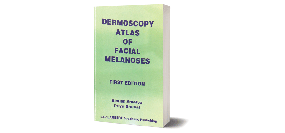
“Dermoscopy Atlas of Facial Melanosis” authored by budding dermatologists Bibush Amatya and Priya Bhusal, introduces readers, intended dermatologists and dermatology residents, to clinical description, dermoscopic features and well labeled, good quality dermoscopic images of various skin conditions manifesting as facial pigmentation.
Facial pigmentation or melanosis is something dermatologists encounter commonly in their day to day practice. In general population, it is a commonplace to find people, especially, young and middle aged, and females to be fretful of the lack of uniformity in facial skin tone. Though this usually is less concerning with respect to any symptom like itching or pain, or any association with internal disease, the pigmentation in itself causes much distress to the afflicted persons regarding his/her appearance.
To the treating physician, facial pigmentation poses challenge in determining the exact nature of pigmentation, as there is a significant clinical overlap between various conditions. This is where dermoscopy comes in handy.
Dermoscope or dermatoscope is a non-invasive tool which helps to visualize deeper skin structures and patterns in higher magnification which are not visible to unaided eyes. Dermoscope is being used across the world, especially to screen suspicious moles in white population.
It is only recently that dermoscope is being utilized in clinical setup by dermatologists in Indian subcontinent and is being considered as the dermatologist’s sthethoscope. Scientific literature regarding usage of dermoscopy in Asian skin is gradually accumulating, mostly from Indian authors and it is a matter of immense pride to see young Nepalese authors like Amatya and Bhusal make a significant contribution in the form of a color atlas.
The book comes in a concise format with 18 chapters, each chapter describing the clinical picture of a type of facial melanosis, its dermoscopic pattern followed by one or more relevant dermoscopic images which are properly labeled and different features marked in the images to enhance clarity for its readers. The descriptions are short and informative as is expected in an atlas.
Images of normal magnification are of excellent quality; however, some images which are magnified to visualize finer patterns have less satisfactory resolution. The language is simple and one can go through the book in an hour or so, if one is well versed with the terms and patterns seen in dermoscopy.
Further understanding can be gained and knowledge consolidated by the practitioner by going through the individual chapters and referring to the images during dermoscopic examination of the patient with particular type of facial pigmentation.
The strengths of the book lies in the fact that it ventures into the less described facial melanoses in the western literature like clofazamine induced pigmentation and peribuccal pigmentation of Brocq, it has an easily readable format and that effort has been made to include the references in each chapter which increases the validity of the content.
Some areas of improvement include image quality in magnified pictures, mentioning exact details of the instrument used and magnification of image, and inclusion of conditions like seborrhoeic melanoses, which is not so rare in our population and lentigo maligna, which, though thankfully rare in our part of the world, holds great importance in terms of early diagnosis and management.
To summarize, the book delivers what is expected out of it and the sincere effort of authors to share their knowledge is appreciable. I believe it will help dermatologists and dermatology residents make better clinical judgments regarding facial pigmentation and at the same time generate more interest in utilizing dermoscopy in their clinical practice.
(The author is associated with Department of Dermatology and Venereology, Nepal Medical College)
Recent News
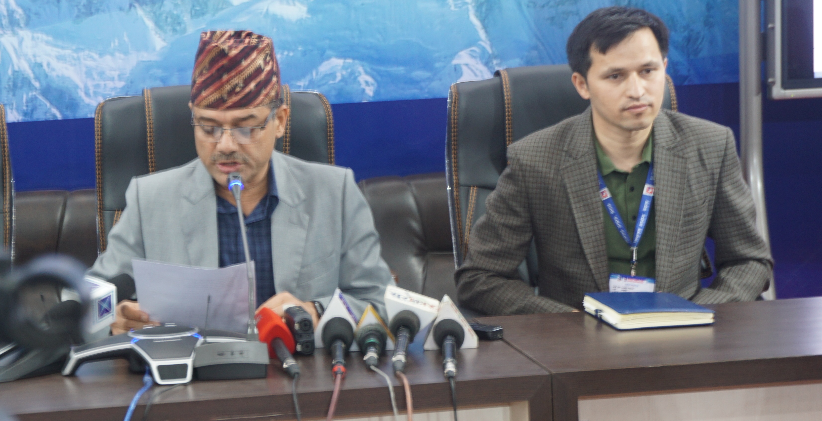
Do not make expressions casting dout on election: EC
14 Apr, 2022
CM Bhatta says may New Year 2079 BS inspire positive thinking
14 Apr, 2022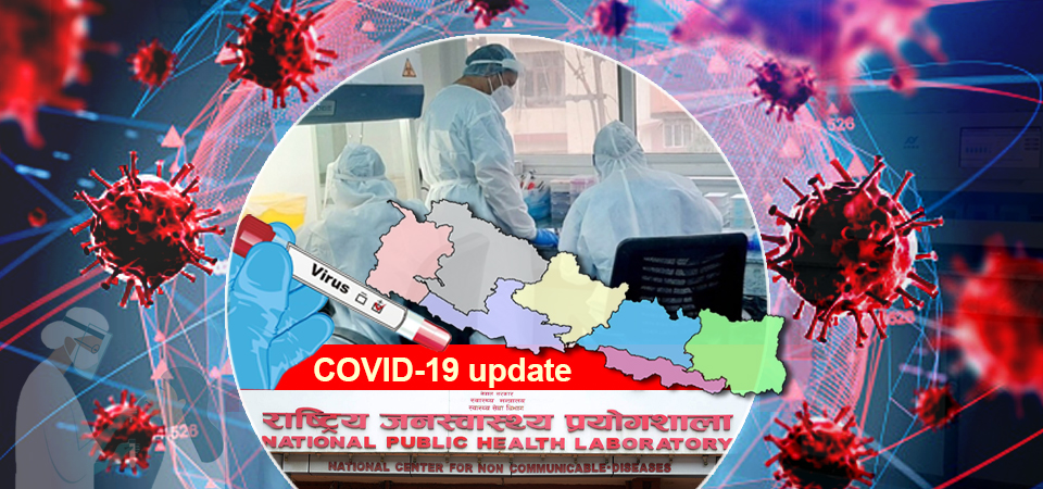
Three new cases, 44 recoveries in 24 hours
14 Apr, 2022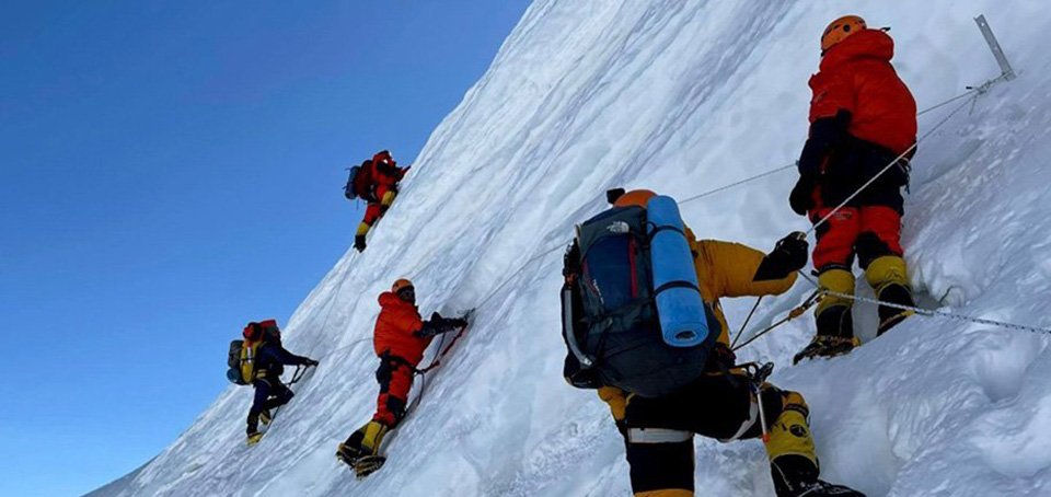
689 climbers of 84 teams so far acquire permits for climbing various peaks this spring season
14 Apr, 2022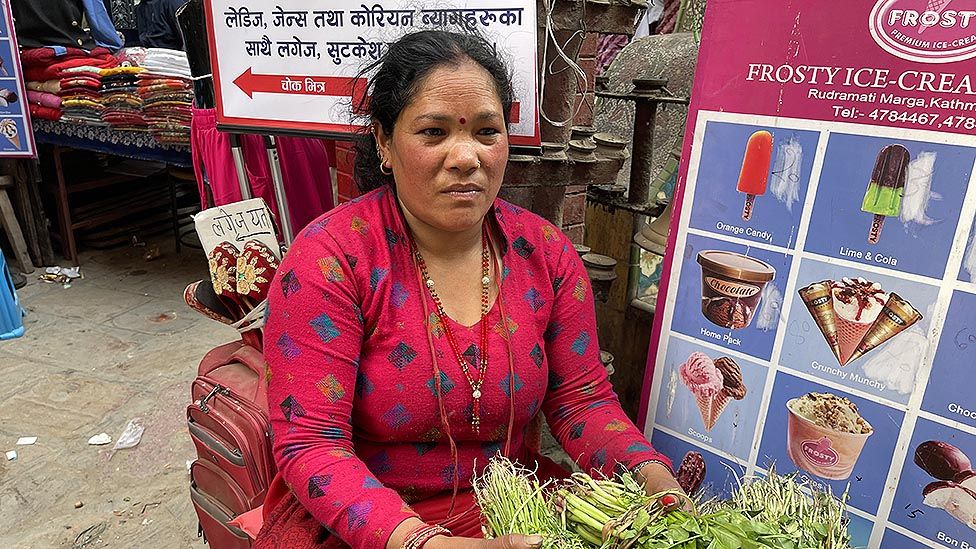
How the rising cost of living crisis is impacting Nepal
14 Apr, 2022
US military confirms an interstellar meteor collided with Earth
14 Apr, 2022
Valneva Covid vaccine approved for use in UK
14 Apr, 2022
Chair Prachanda highlights need of unity among Maoist, Communist forces
14 Apr, 2022
Ranbir Kapoor and Alia Bhatt: Bollywood toasts star couple on wedding
14 Apr, 2022
President Bhandari confers decorations (Photo Feature)
14 Apr, 2022

